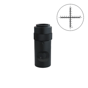
Quick Overview
Video Zoom Lens Compatible. Coupler Magnification: 1X. Cross Line. Reticle Dimensions: Dia. 16x1.5mm. Background Type: Positive. For MZ0201 Series Video Zoom Body. For Reticle Size: 16mm.
MZ02016411 Reticle Coupler
Coupler/C-mount Adapter
| Coupler Mount Type for Body | Thread Screw |
| Coupler Mount Size for Body | Dia. 25.4x1/36 in. |
| Adjustable Coupler | Adjustable |
| Coupler for Microscope Type | Video Zoom Lens Compatible |
| Coupler Magnification | 1X |
| C/CS-Mount Coupler | C-Mount |
| Reticle Type | Cross Line |
| Reticle Dimensions | Dia. 16x1.5mm |
| Surface Treatment | Electroplating Black |
| Material | Metal |
| Color | Black |
| Net Weight | 0.12kg (0.26lbs) |
| Applied Field | For MZ0201 Series Video Zoom Body |
| For Reticle Size | 16mm |
Reticle
| Cross Line Reticle ( Dia. 16mm) | |
| Reticle Type | Cross Line |
| Reticle Dimensions | Dia. 16x1.5mm |
| Diameter Tolerance | -0.02一 -0.07mm |
| The Maximum Tolerance of The Center Offset Is | 0.05一0.1mm |
| Reticle Coating Type | Chrome Plated |
| Background Type | Positive |
| Net Weight | 0.002kg (0.004lbs) |
Technical Info
Instructions
Coupler and Camera AdapterClose Λ
| Coupler or camera adapter, is also called photo eyepiece, CCTV adapter, CCD adapter, camera adapter, digital camera adapter, and camera adapter etc. Coupler is an optical imaging lens that uses mechanical adapter device to connect the camera to the microscope, and project the image onto the camera's target through the adapter. Coupler/C-mount adapters have different magnifications to capture images of different magnifications and field of view. In optical imaging, optical parameters such as chromatic aberration, distortion, and field curvature are also to be considered to ensure image quality. At the same time, for some other couple/C-mount adapters, factors such as the position and angle of the camera are also considered to suit the needs of the observational environment. |
Coupler/C-mount AdapterClose Λ
| Coupler/C-mount adapter is an adapter commonly used for connection between the C-adapter camera (industrial camera) and a microscope. |
Adjustable CouplerClose Λ
| On the coupler/C-mount-adapter, there is an adjustable device to adjust the focal length. |
Coupler for Microscope TypeClose Λ
| Different coupler/C-mount-adapters are suitable for different microscopes. For some, some adapter accessories need to be replaced. See the applicable range of each coupler/C-mount-adapter for details. |
Coupler MagnificationClose Λ
| Coupler magnification refers to the line field magnification of the coupler/C-mount-adapter. With different magnifications of the adapter lens, images of different magnifications and fields of view can be obtained. The size of the image field of view is related to the sensor size and the coupler/C-mount-adapter magnification. Camera image field of view (mm) = sensor diagonal / coupler/C-mount-adapter magnification. For example: 1/2 inch sensor size, 0.5X coupler/C-mount-adapter coupler, field of view FOV (mm) = 8mm / 0.5 = 16mm. The field of view number of the microscope 10X eyepiece is usually designed to be 18, 20, 22, 23mm, less than 1 inch (25.4mm). Since most commonly used camera sensor sizes are 1/3 and 1/2 inches, this makes the image field of view on the display always smaller than the field of view of the eyepiece for observation, and the visual perception becomes inconsistent when simultaneously viewed on both the eyepiece and the display. If it is changed to a 0.5X coupler/C-mount-adapter, the microscope image magnification is reduced by 1/2 and the field of view is doubled, then the image captured by the camera will be close to the range observed in the eyepiece. Some adapters are designed without a lens, and their optical magnification is considered 1X. |
C/CS-Mount CouplerClose Λ
| At present, the coupler/C-mount adapter generally adopts the C/CS-Mount adapter to match with the industrial camera. For details, please refer to "Camera Lens Mount". |
ReticleClose Λ
| Reticle is generally also referred to as eyepiece reticle, or reticule, graticule, cross hair. Reticle is an optical component with a certain mark placed inside the eyepiece. Based on different applications, reticle can be used for measurement, calibration or aiming. Reticle is mainly used for the measurement of length, angle or area of the object to be measured under the microscope. The reticle measurement is a "non-contact measurement", that is, the measurement value is obtained by measuring the optical image without touching the object to be measured, which is very suitable for some small specimens, organisms, and irregular objects. Eyepiece reticle has patterns of various shapes and sizes. Common types of eyepiece reticle are: straight, cross, mesh, circle, angle or combination shape. Between each grid it is also equidistant. However, for eyepiece reticle, one cannot read directly the number under the microscope, but convert firstly the multiple after magnification of the microscope objective lens. In short, after the object is being magnified by the objective lens, the real image of the object reaches the focal length of the eyepiece (10 mm below the fixed surface of the eyepiece), which is exactly the position of the eyepiece reticle, and what the eyepiece reticle reads is actually the image of the object after being magnified by the objective lens. Therefore, for the actual numerical value, the actual size of the image should be divided by the magnification of the objective lens. In addition, for eyepiece reticle measurement, it can also be calibrated first by the objective micrometer before measurement. The method is: first, place an objective micrometer on the stage, after the focus is clear, record the magnification number of the objective. Then, the eyepiece reticle is overlapped with the scale pattern of the objective micrometer, so that the 0 points of the two are aligned, a scale value with a completely coincident scale is found backward, the grid values of the reticle eyepiece and the objective micrometer are respectively read and converted, and then the calibration value is used as the actual measurement value of the eyepiece reticle. This method is relatively more complicated. First, it is necessary to constantly convert the reading value and the calibration value of the eyepiece. Secondly, each time when the objective lens with different magnifications for observation is changed, it needs to be re-calibrated. This is only suitable for use in strongly repetitive microscope observations and work in order to be efficient. Reticle Installation The reticle is installed in the eyepiece tube, and some eyepieces have been installed with reticle before leaving the factory. Since the requirements are different, users can also buy different reticle, and then install it on their own microscope. To install the reticle yourself, first make sure that the eyepiece of the microscope can be self-removed from the microscope eyepiece tube (generally, for microscopes, all their eyepieces can be removed, and some need to loosen the screws fastened on the microscope eyepiece tube to remove the eyepiece.) For eyepieces on which reticle can be installed, you should pay attention to the following features and requirements: 1. Whether the tube wall of the eyepiece has a “mounting/installation surface” on which the reticle is placed. Generally, the eyepieces are located 10mm below the lower lens. This position is the focal plane of the eyepiece. The reticle is installed in this position to be clear. 2. Whether it has "Eyepiece Reticle Fix Ring". There are generally two ways for this fix ring: one is that there is the thread on the inner wall of the eyepiece tube, a metal fix ring with a card slot for positioning when using a screwdriver, by rotating the screwdriver, the reticle is pressed on the inner wall of the eyepiece. There is also a"plug ring type"fix ring, usually made of plastic material, which is elastic and inserted into the eyepiece tube, and then stuck on the inner wall of the eyepiece tube to press the reticle. If this"fix ring" is missing in the eyepiece tube, please contact your service provider to describe the above situation, and some service providers can provide this fix ring. 3. The tick marks of the reticle are all on top of the reticle. Generally, all reticles of the glass material have a certain thickness, and the tick marks of the reticle is on top of the reticle to ensure that all the tick marks are in the eyepiece focal plane (10 mm below the eyepiece) when using the reticle of different thickness. 4. Measure the diameter of the inner wall of the microscope eyepiece tube, to select the appropriate size of the reticle. Upon understanding the above, if you need to choose reticle for different purpose of use, please visit Bolioptics.com to select reticle with a different pattern for use. |
PackagingClose Λ
| After unpacking, carefully inspect the various random accessories and parts in the package to avoid omissions. In order to save space and ensure safety of components, some components will be placed outside the inner packaging box, so be careful of their inspection. For special packaging, it is generally after opening the box, all packaging boxes, protective foam, plastic bags should be kept for a period of time. If there is a problem during the return period, you can return or exchange the original. After the return period (usually 10-30 days, according to the manufacturer’s Instruction of Terms of Service), these packaging boxes may be disposed of if there is no problem. |
Optical Data
| Camera Image Sensor Specifications | |||
| No. | Camera Image Sensor Size | Camera image Sensor Diagonal | |
| (mm) | (inch) | ||
| 1 | 1/4 in. | 4mm | 0.157" |
| 2 | 1/3 in. | 6mm | 0.236" |
| 3 | 1/2.8 in. | 6.592mm | 0.260" |
| 4 | 1/2.86 in. | 6.592mm | 0.260" |
| 5 | 1/2.7 in. | 6.718mm | 0.264" |
| 6 | 1/2.5 in. | 7.182mm | 0.283" |
| 7 | 1/2.3 in. | 7.7mm | 0.303" |
| 8 | 1/2.33 in. | 7.7mm | 0.303" |
| 9 | 1/2 in. | 8mm | 0.315" |
| 10 | 1/1.9 in. | 8.933mm | 0.352" |
| 11 | 1/1.8 in. | 8.933mm | 0.352" |
| 12 | 1/1.7 in. | 9.5mm | 0.374" |
| 13 | 2/3 in. | 11mm | 0.433" |
| 14 | 1/1.2 in. | 12.778mm | 0.503" |
| 15 | 1 in. | 16mm | 0.629" |
| 16 | 1/1.1 in. | 17.475mm | 0.688" |
| Digital Magnification Data Sheet | ||
| Image Sensor Size | Image Sensor Diagonal size | Monitor |
| Screen Size (24in) | ||
| Digital Zoom Function | ||
| 1/3 in. | 6mm | 101.6 |
| 1. Digital Zoom Function= (Screen Size * 25.4) / Image Sensor Diagonal size | ||
| Contains | ||||||||||
| Parts Including | ||||||||||
| ||||||||||
| Packing | |
| Packaging Type | Carton Packaging |
| Packaging Material | Corrugated Carton |
| Packaging Dimensions(1) | 7.5x4x11.5cm (2.953x1.575x4.528″) |
| Inner Packing Material | Plastic Bag |
| Ancillary Packaging Materials | Styrofoam |
| Gross Weight | 0.14kg (0.31lbs) |
| Minimum Packaging Quantity | 1pc |
| Transportation Carton | Carton Packaging |
| Transportation Carton Material | Corrugated Carton |
| Transportation Carton Dimensions(1) | 15.2x15.2x15.2cm (6x6x6″) |
| Total Gross Weight of Transportation(kilogram) | 0.27 |
| Total Gross Weight of Transportation(pound) | 0.60 |


