Quick Overview
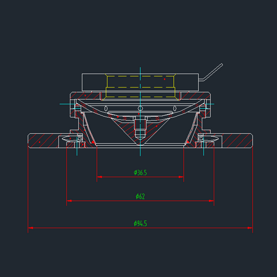
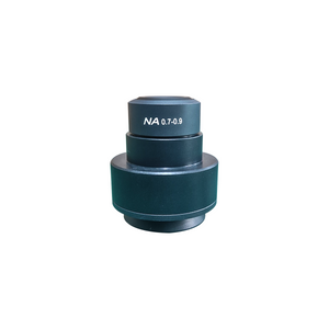
Darkfield Attachment Type: Dark Field. Darkfield Attachment Mount Size: Dia. 95x5mm.
JM12062201 Darkfield Condenser for Stereo Microscope
Darkfield Attachment
| Darkfield Attachment Type | Dark Field |
| Darkfield Attachment Mount Size | Dia. 95x5mm |
| Surface Treatment | Electroplating Black |
| Material | Aluminum |
| Color | Black |
| Net Weight | 0.14kg (0.30lbs) |
Technical Info
Instructions
IlluminatorClose Λ
| The conditions of different illumination of the microscope are a very important parameter. Choosing the correct illumination method can improve the resolution and contrast of the image, which is very important for observing the imaging of different objects. The wavelength of the light source is the most important factor affecting the resolution of the microscope. The wavelength of the light source must be smaller than the distance between the two points to be observed in order to be distinguished by the human eye. The resolution of the microscope is inversely proportional to the wavelength of the light source. Within the range of the visible light, the violet wavelength is the shortest, providing also the highest resolution. The wavelength of visible light is between 380~780nm, the maximum multiple of optical magnification is 1000-2000X, and the limit resolution of optical microscope is about 200nms. In order to be able to observe a much smaller object and increase the resolution of the microscope, it is necessary to use light having a much shorter wavelength as the light source. The most commonly used technical parameters for describing illumination are luminescence intensity and color temperature. Luminescence intensity, with lumen as unit, is the physical unit of luminous flux. The more lumens, the stronger the illumination. Color temperature, with K (Kelvin) as unit, is a unit of measure indicating the color component of the light. The color temperature of red is the lowest, then orange, yellow, white, and blue, all gradually increased, with the color temperature of blue being the highest. The light color of the incandescent lamp is warm white, its color temperature is 2700K, the color temperature of the halogen lamp is about 3000K, and the color temperature of the daylight fluorescent lamp is 6000K. A complex and complete lighting system can include a light source, a lampshade or lamp compartment, a condenser lens, a diaphragm, a variety of wavelength filters, a heat sink cooling system, a power supply, and a dimming device etc. Select and use different parts as needed. Of which, selection and use of the illuminating light source is the most important part of the microscope illumination system, as and other components are designed around the illuminating wavelength curve and characteristics of the illuminating light source. Some of the microscope light sources are pre-installed on the body or frame of the microscope, and some are independent. There are many types and shapes of light sources. Depending on the requirements of the microscope and the object to be observed, one type or multiple types of illumination at the same time can be selected. In addition, the whole beam and band adjustment of the light source, the position and illumination angle of the light source, and the intensity and brightness of the light all have a great influence on the imaging. For microscope imaging, a good lighting system may be a system that allows for more freedom of adjustment. In actual work, such as industry, too many adjustment mechanisms may affect the efficiency of use, therefore choose the appropriated configured lighting conditions is very important. |
Dark FieldClose Λ
| Bright field illumination is that direct light shining on the object to be observed and the background enters directly into the objective lens, producing a background of a bright image; the dark field corresponds to the bright field, so that the light is obliquely irradiated onto the surface of the sample, and the direct light is not allowed to pass through the aperture of the objective lens, so as to be able to observe object details in a black background. The light source adopts parallel light, and an annular visor is placed at an appropriate position on the incident light path to cover the central portion of the light. The surrounding light passing through the annular visor is a hollow cylindrical beam and is incident on the vertical illuminator. The vertical illuminator of the dark field illumination is a circular reflector that reflects the cylindrical beam upwards and projects it onto the reflector of the condenser, so that the reflected light is concentrated on the surface of the sample. Since the reflected light is highly tilted, the light cannot enter the objective lens, therefore the field of view is dark. Only in the concave place of the observed object can light enter the objective lens, and the bright white image is reflected in the dark field of view, called "dark field of view " (dark field) illumination. In most cases, the bright portion of the black-and-white image obtained by dark field illumination is considered to be the black portion of the black-and-white image obtained by bright field illumination. Dark field illumination improves the contrast of the image, making the color of the image natural and uniform. The angle of the incident beam is extremely large, which increases the effective numerical aperture of the objective lens, and the ability to identify details is higher than that of bright field illumination. Dark field illumination must ensure that the system is clean, as dust particles can form bright spots in the dark field, which can affect the results of observation. |
PackagingClose Λ
| After unpacking, carefully inspect the various random accessories and parts in the package to avoid omissions. In order to save space and ensure safety of components, some components will be placed outside the inner packaging box, so be careful of their inspection. For special packaging, it is generally after opening the box, all packaging boxes, protective foam, plastic bags should be kept for a period of time. If there is a problem during the return period, you can return or exchange the original. After the return period (usually 10-30 days, according to the manufacturer’s Instruction of Terms of Service), these packaging boxes may be disposed of if there is no problem. |
| Packing | |
| Inner Packing Material | Plastic Bag |
| Gross Weight | 0.28kg (0.62lbs) |
| Minimum Packaging Quantity | 1pc |
| Transportation Carton | Carton Packaging |
| Transportation Carton Material | Corrugated Carton |
| Transportation Carton Dimensions(1) | 15.2x15.2x15.2cm (6x6x6″) |
| Total Gross Weight of Transportation(kilogram) | 0.28 |
| Total Gross Weight of Transportation(pound) | 0.62 |


